Wound Care Basics
Wound Management: How Specialists Measure Healing Progress
Discover how specialists track wound management progress visit to visit through precise measurements, tissue analysis, tunneling checks, and skin health.
Published · Anchor Wound Management
Overview
- Clinical wound tracking relies on objective dimensions, including length, width, and depth, to evaluate tissue changes rather than relying on casual visual impressions.
- Specialists map internal changes like undermining and tunneling using a clock-face orientation to verify healing from deep tissue outward.
- Documenting tissue types, exudate volume, and the surrounding periwound skin helps providers spot infection or stalled repair early.
- Tracking quantitative data visit to visit ensures therapies are adjusted promptly if an injury fails to progress over consecutive weeks.
Healing is rarely straightforward for individuals managing complex injuries. Effective wound management requires far more than simply replacing a bandage and checking whether an injury looks better to the naked eye. In clinical practice, measuring recovery is an exact science that translates physical changes into actionable clinical data. Whether evaluations take place in an outpatient clinic or through mobile bedside wound care visits in Dallas–Fort Worth, providers utilize systematic benchmarks to monitor every stage of tissue repair.
Without standardized measurements, subtle indicators of stagnation or deterioration can easily be overlooked. A sore might appear smaller on the surface while quietly expanding beneath intact skin, or changes in drainage might signal complications before severe discomfort arises. By documenting physical characteristics at every appointment, clinicians establish an objective record that guides ongoing clinical decisions.
Standard Metrics Used in Clinical Wound Management
Before any therapy begins, a specialist must identify the underlying etiology, anatomical location, and specific classification of the wound. According to clinical wound assessment guidelines published in StatPearls, comprehensive assessment protocols evaluate the initial cause, exact location, and tissue type, followed by ongoing measurements of length, width, depth, undermining, tunneling, wound bed composition, exudate, periwound condition, and signs of infection at every subsequent visit to track healing trajectories.
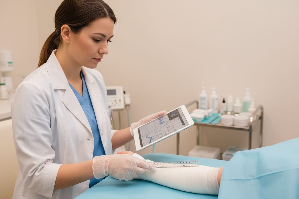
The most fundamental components of this evaluation are the three linear dimensions: length, width, and depth. To ensure consistency across different clinicians and appointments, providers follow the clock method of measurement. In this system, the patient’s head represents 12:00, while the feet represent 6:00. The greatest distance along this axis is recorded as length, while width is measured perpendicular from 9:00 to 3:00. Measuring instruments typically include disposable metric rulers calibrated in millimeters or centimeters.
Depth presents a unique clinical challenge because wounds rarely display a flat floor. Clinicians assess depth by gently placing a sterile, cotton-tipped probe into the deepest visible area of the wound bed, noting the level even with surrounding skin margins, and measuring that distance against a metric ruler. Calculating surface area and tracking depth over time gives the care team quantifiable evidence showing whether the injury is contracting appropriately.
Detecting Hidden Space: Tunneling and Undermining
A wound cannot be fully evaluated solely by its visible surface margins. Two complications that often distort standard surface measurements are undermining and tunneling. Undermining occurs when tissue destruction extends laterally beneath intact skin edges along the wound perimeter. This creates an overhanging shelf of skin, meaning the true anatomical boundary is substantially larger than the surface opening suggests.
Tunneling involves narrow channels extending from wound margins into deeper subcutaneous tissue or muscle. Both conditions result from shear forces, pressure, or persistent infection. Left undetected, these hidden spaces collect fluid, foster bacterial growth, and prevent the wound from closing from the base upward.
To assess these features, clinicians gently probe the perimeter with a sterile cotton swab, documenting location and depth using the clock-face reference. For example, a specialist might record two centimeters of undermining from 1:00 to 4:00, or a three-centimeter tunnel at 7:00. Tracking these hidden spaces visit to visit is critical; a decrease in tunnel depth demonstrates genuine healing even if external surface dimensions remain identical.
Analyzing the Wound Bed: Granulation, Slough, and Eschar
Beyond dimensions, specialists evaluate biological activity within the wound bed. Healing progress is deeply linked to the specific phases of tissue recovery, which manifest as distinct tissue types. Clinicians typically estimate and record the percentage of each tissue type visible across the wound base during every examination.
- Granulation Tissue: This is healthy, highly vascularized connective tissue that fills the wound space. It presents as moist, bumpy, and bright beefy red or deep pink. A rising percentage of granulation indicates robust microcirculation and active tissue regeneration.
- Slough: Slough consists of non-viable cellular debris, fibrin, and bacteria. It appears moist, stringy, or mucinous, ranging in color from soft yellow and cream to grayish-tan. Slough prevents healthy granulation from forming and must be cleared through clinical debridement.
- Eschar: Eschar represents dead, necrotic tissue that has hardened into a dry, leathery, black or dark brown crust. While stable, dry eschar on a heel may sometimes be monitored, wet eschar traps underlying infection and halts closure.
- Epithelial Tissue: During final recovery stages, new epithelial cells migrate across the granulation bed from edges, appearing as pale pink, thin margins signaling closure.
When clinical tracking shows slough or eschar decreasing while viable granulation increases, the wound is progressing favorably toward re-epithelialization.
Evaluating Exudate and Periwound Skin Health
The fluid produced by a wound, known as exudate, offers direct clues regarding inflammation and microbial balance. Clinicians categorize drainage by volume and character. Serous drainage is clear or pale amber and watery, reflecting normal inflammatory processes. Sanguineous fluid is thin and red due to fresh blood, while serosanguineous fluid is light pink. In contrast, purulent drainage is thick, opaque, yellow, green, or milky, often carrying an unpleasant odor that suggests bacterial colonization or infection.
Equally critical is the condition of the periwound skin—the area extending at least four centimeters beyond the wound edges. Healthy margins support cellular migration, but damaged skin compromises healing. Clinicians examine this area for maceration (skin turned white, soft, and fragile from excessive moisture), erythema (redness indicating inflammation or infection), induration (firm, hardened tissue caused by edema or deep infection), and denudation (loss of the superficial epidermal layer). Tracking periwound health ensures that dressing selections properly manage fluid without compromising healthy surrounding tissue.
Managing Chronic Wounds and Underlying Conditions
When normal cellular repair processes fail to advance, specialized intervention becomes essential. As detailed in research on chronic wound management from InformedHealth.org, a wound is considered chronic if it has not started to heal after four to twelve weeks despite treatment. These persistent wounds usually stem from poor circulation, diabetes, or a weakened immune system. Evidence-based care requires cleaning and appropriate dressings, debridement of dead tissue, and consideration of negative pressure wound therapy or skin grafts when healing stalls. Clinicians must also treat underlying medical conditions while providing pain relief and psychological support.
Patients dealing with systemic diseases that impair healing frequently require these multidisciplinary approaches. Visit-to-visit tracking provides the objective threshold for adjusting treatments. If detailed records show no reduction in surface area or continuous devitalized tissue over several appointments, clinicians know immediately that the current management strategy must be recalibrated.
Warning Signs Requiring Prompt Evaluation
Patients and caregivers should never attempt self-debridement or manage worsening symptoms alone. As emphasized in clinical wound guidelines, signs of advancing infection require immediate clinical intervention. Seek urgent medical attention if you experience spreading redness, escalating pain, new foul odor, increased heat around the site, sudden heavy or purulent drainage, or systemic red flags such as fever and chills.
Next Steps in Your Care
Consistent clinical tracking eliminates guesswork. If you need specialized guidance, please contact Anchor Wound Management to arrange an evaluation at our clinics or through bedside care.
Sources
- StatPearls. Wound Assessment (StatPearls). 2023.
- InformedHealth.org. Chronic wounds: What are the treatment options for chronic wounds?. 2025.