Conditions
Diabetic Leg Ulcer Treatment: Why the Wound Location Matters
Explore how wound position guides diabetic leg ulcer treatment. Understand neuropathic, arterial, and venous ulcers to support timely medical intervention.
Published · Anchor Wound Management

Overview
- The physical location of a lower extremity break in the skin provides crucial diagnostic clues about underlying vascular and nerve health.
- Plantar wounds on the bottom of the foot typically point to peripheral neuropathy, requiring specialized pressure relief strategies.
- Wounds on toes or the posterior heel often indicate compromised arterial blood flow that demands vascular evaluation before interventions begin.
- Sores in the lower leg gaiter area usually reflect chronic venous insufficiency, which requires careful assessment before compression therapy can start.
Navigating diabetic leg ulcer treatment can feel overwhelming, especially when a small sore appears unexpectedly and refuses to heal. For individuals living with diabetes, lower extremity skin breakdowns are not all the same. While high blood sugar is a common denominator, the precise anatomical position of an ulcer provides essential diagnostic clues regarding what is happening beneath the skin. Whether an open sore develops under the ball of the foot, along the tip of a toe, or higher up around the ankle, its location points directly toward the underlying physiological failure driving the breakdown.
According to the National Institute of Diabetes and Digestive and Kidney Diseases guidance on foot problems, daily foot inspections are critical because nerve damage and reduced blood flow significantly raise the risk of serious complications. Recognizing why an ulcer appeared in a specific spot allows clinicians to tailor treatment plans to the actual root cause rather than merely applying topical bandages to surface symptoms.
How Wound Location Guides Diabetic Leg Ulcer Treatment
Clinicians view the lower limb as a map divided into distinct vascular and neurological territories. When assessing an open sore, wound care specialists evaluate three primary forces: mechanical stress combined with loss of sensation, arterial insufficiency resulting in tissue starvation, and venous hypertension causing fluid pooling. A patient living with diabetes can develop any of these issues, and in many cases, multiple pathologies coexist.
Understanding these distinct anatomical zones helps determine whether a patient requires pressure redistribution, revascularization to restore blood flow, or edema management to reduce fluid buildup. Selecting the correct therapeutic approach is essential because applying the wrong intervention can cause harm. For instance, applying tight compression wraps to an ulcer caused by poor arterial flow can completely cut off blood supply to the limb. Therefore, observing the anatomical location is the initial foundational step in establishing an accurate clinical diagnosis and creating an individualized plan of care.
Plantar Surface and Pressure Points: Neuropathic Ulcers
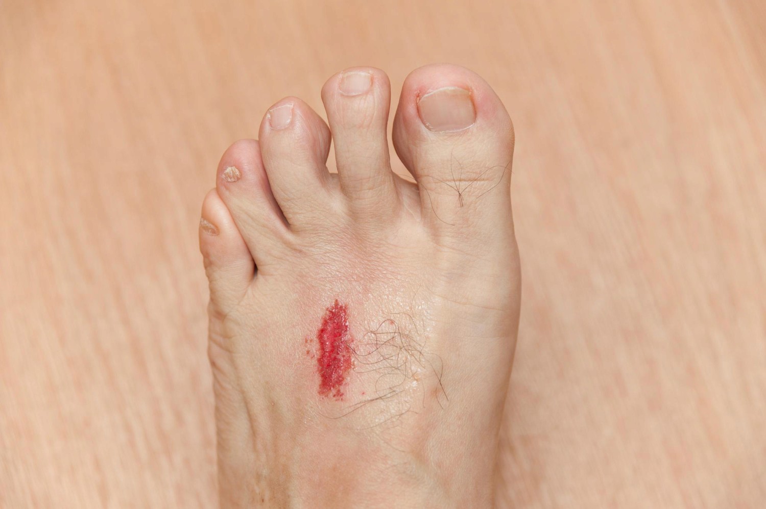
The bottom of the foot, known medically as the plantar surface, bears the immense mechanical burden of daily walking. Common sites for breakdown include the metatarsal heads under the ball of the foot, the base of the big toe, and beneath the heel pad. When a non-healing sore appears in these weight-bearing zones, it is overwhelmingly classified as a neuropathic ulcer.
Diabetic peripheral neuropathy gradually dulls or eliminates the protective sensation that normally alerts a person to pain, friction, or pressure. When patients lose this sensation, repetitive trauma goes completely unnoticed. A tiny pebble in a shoe, a rigid internal seam, or uneven pressure caused by natural foot deformities like bunions or hammertoes can wear through the dermis over hours of walking. The body often attempts to protect itself by forming a thick callus, but continued pressure turns that callus into an internal blister that eventually breaks open into an ulceration.
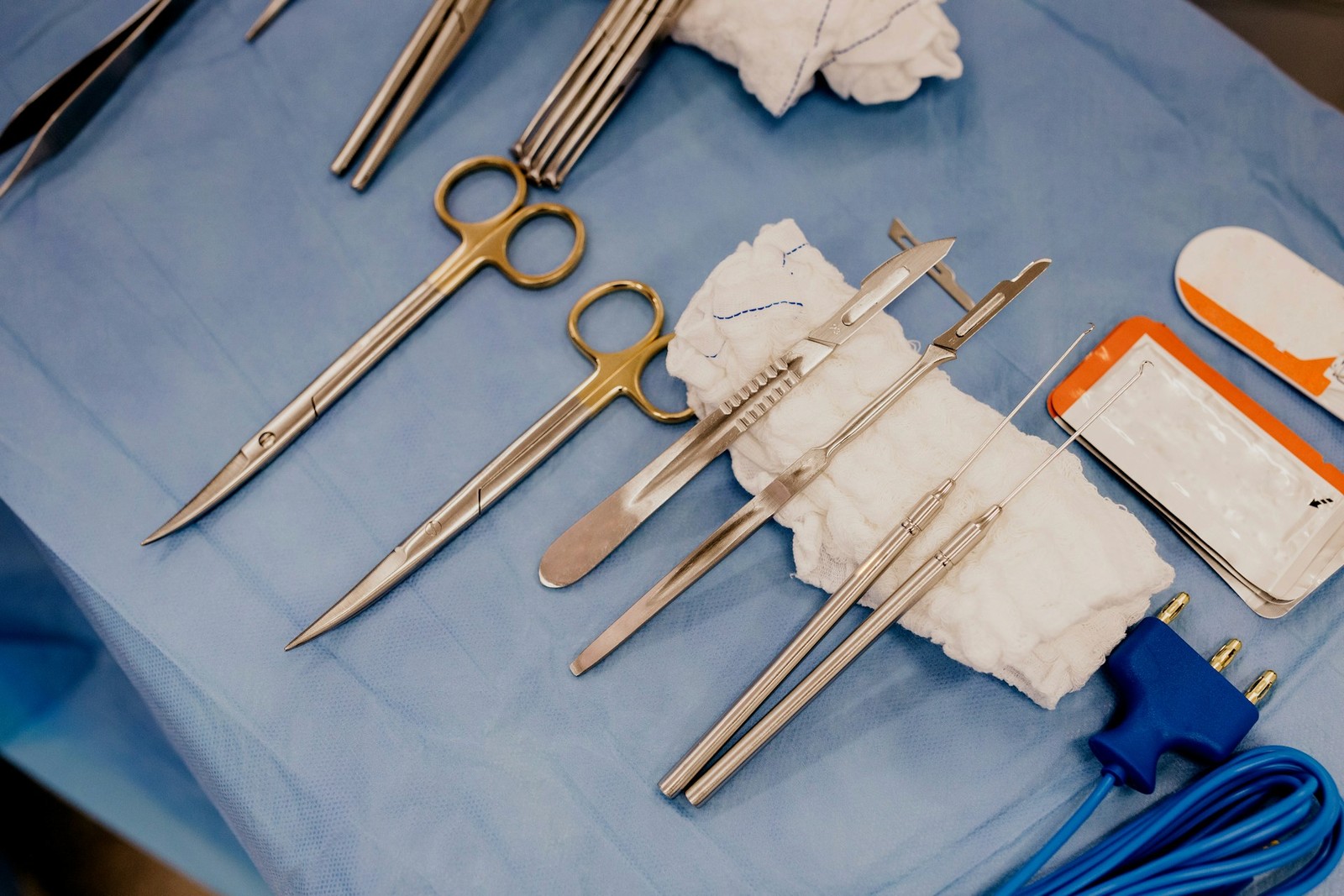
Addressing these sores requires specialized comprehensive care for diabetic foot ulcers. The primary intervention for plantar ulcers is offloading, which means redistributing weight away from the wound bed through specialized footwear, cast boots, or custom orthotics. Additionally, clinicians frequently perform selective sharp debridement to remove non-viable, calloused tissue and bacterial biofilm from the wound edges. By removing dead tissue and relieving persistent pressure, the body is given an opportunity to generate healthy granulation tissue.
Toes and Heels: Arterial Ulcers and Ischemia
In contrast to plantar surfaces, ulcers that develop on the tips of the toes, between adjacent toes, on the lateral borders of the foot, or over the posterior calcaneus (heel bone) often signal an arterial problem. These areas represent the furthest points from the heart and are particularly vulnerable when blood vessels narrow.
Peripheral artery disease restricts the delivery of oxygenated blood, nutrients, and immune cells to the furthest extremities. When arterial inflow is compromised, even trivial friction from snug shoes or resting the heels against a bed mattress can trigger rapid tissue necrosis. Arterial ulcers typically present with a "punched-out" appearance, pale or gray wound beds, minimal drainage, and profound localized pain that often worsens when the legs are elevated. The surrounding skin may appear shiny, hairless, cool to the touch, and thin.
The IWGDF guidelines on diabetes-related foot disease emphasize that managing suspected arterial ulcers requires thorough vascular assessment and multidisciplinary coordination. Clinicians must confirm adequate blood flow before initiating aggressive local wound therapies. If blood supply is severely restricted, specialized vascular procedures to restore circulation must take precedence. Debridement is often deferred on dry, stable arterial eschars until vascular specialists can evaluate whether the tissue has sufficient blood supply to heal, preventing further tissue loss.
The Gaiter Area: Venous Ulcers in Diabetic Patients
The lower leg—specifically the "gaiter area" extending from just below the calf muscle down to the ankle bones—is the typical home of venous ulcers. While diabetes is primarily associated with foot ulcers, many older individuals with diabetes also suffer from chronic venous insufficiency, leading to complex mixed-etiology wounds.
As documented in clinical reviews of venous leg ulcers published in StatPearls, these lesions represent a late manifestation of chronic venous hypertension, classified clinically as CEAP C6 active ulcers. Faulty valves inside the leg veins fail to pump deoxygenated blood back up toward the heart effectively. Blood pools in the lower legs, elevating pressure within the microvasculature. This sustained venous hypertension causes red blood cells to leak into surrounding skin tissues, resulting in characteristic brown or rust-colored staining known as hemosiderin hyperpigmentation. Eventually, the congested, swollen skin becomes fragile, inflamed, and breaks down into shallow, irregularly shaped wounds with heavy fluid drainage.
Compression therapy serves as the clinical cornerstone for managing venous hypertension, but diabetic patients require extra caution. Because diabetes frequently causes silent peripheral artery disease, clinicians must carefully measure the ankle-brachial index before applying compression wraps. Applying high-pressure compression wraps to a leg with undiagnosed arterial blockage can damage compromised tissue. Professional evaluation ensures that compression levels are calibrated safely to patient physiology.
Recognizing Complications: When to Seek Urgent Medical Care
Regardless of wound location, individuals living with diabetes must monitor lower extremity sores closely. Impaired immunity can cause minor ulcers to deteriorate rapidly into deep tissue infections. Patients and caregivers should review the distinctions detailed in our guide on identifying diabetic wound red flags to recognize complications early.
Seek immediate medical evaluation if you encounter spreading redness, foul-smelling drainage, fever, chills, black tissue discoloration, or exposed tendon and bone.
Comprehensive Wound Care Across Dallas–Fort Worth
Because every ulcer location reflects a distinct underlying cause, successful recovery depends on an accurate clinical diagnosis and tailored therapy. Anchor Wound Management provides advanced evaluations both in our outpatient clinics and directly at the bedside through our mobile care program. If you or a loved one is dealing with a non-healing lower extremity sore, contact Anchor Wound Management to arrange an evaluation.
Sources
- International Working Group on the Diabetic Foot. IWGDF Guidelines on the prevention and management of diabetes-related foot disease (2023 update). 2023.
- National Institute of Diabetes and Digestive and Kidney Diseases (NIDDK). Diabetes & Foot Problems.
- Robles-Tenorio A, Lev-Tov H, Ocampo-Candiani J. Venous Leg Ulcer (StatPearls). 2022.