Conditions
Leg Ulcer Treatment: Why Identifying the Cause Matters
Effective leg ulcer treatment requires identifying the cause. Learn how venous, arterial, and diabetic wounds differ and when to seek expert clinical care.
Published · Anchor Wound Management
Overview
- A lower-leg wound cannot heal effectively until a clinician accurately diagnoses its underlying vascular or physical cause.
- Applying compression wraps to an undiagnosed ulcer is dangerous if underlying arterial blood flow is inadequate.
- Different wound types require distinct medical therapies, such as offloading pressure, surgical revascularization, or specialized dressings.
- Regular clinical monitoring helps identify limb-threatening infections and addresses systemic barriers that halt tissue repair.
Navigating leg ulcer treatment can feel overwhelming, especially when an open sore on the lower calf, ankle, or foot refuses to heal after several weeks. Many people assume that a wound on the lower extremity simply requires standard antiseptic ointment and clean gauze. However, treating a leg break in the skin without understanding why it formed is one of the most common reasons healing stalls. The lower legs endure significant physical demands, carrying body weight while relying on complex vascular networks to circulate oxygenated blood downward and return deoxygenated blood back toward the heart.
When an ulcer appears, it is rarely an isolated skin condition. Instead, it serves as an outward signal of an internal circulatory or mechanical imbalance. Whether the failure stems from venous hypertension, restricted arterial inflow, nerve damage, or sustained physical pressure, matching the intervention to the specific diagnosis is the single most important clinical step. A therapy that helps one type of wound can seriously injure tissue if applied to another.
Why Etiology Governs Leg Ulcer Treatment
Every lower-extremity wound has an origin story. In clinical practice, clinicians categorize chronic wounds by their primary etiology, meaning the root biological problem responsible for tissue breakdown. If a care team attempts treatment without diagnosing this origin, therapies are chosen blindly. Healing requires healthy microcirculation, cellular nutrition, waste removal, and tissue moisture balance. When these physiological elements are compromised, traditional first-aid ointments cannot overcome the cellular deficit.
Furthermore, systemic health challenges frequently overlap to delay tissue repair. Factors such as uncontrolled blood sugar, systemic inflammation, chronic edema, and nutritional deficiencies can halt the cellular repair process altogether. Exploring the broader biological reasons why wounds fail to heal illustrates why superficial skin treatments rarely resolve deep-seated vascular ulcerations without comprehensive clinical oversight.
Venous Ulcers and the Crucial Role of Vascular Screening
Venous insufficiency stands as the most frequent culprit behind chronic lower-leg sores. As documented in a clinical review of venous leg ulcers published by StatPearls, these lesions represent a late-stage manifestation of sustained venous hypertension and chronic venous insufficiency, which occur more frequently in older adults. Within healthy veins, tiny one-way valves keep blood flowing upward against gravity. When these valves weaken, blood pools in the lower extremities, increasing pressure within local capillaries.
According to clinical resources on venous insufficiency from MedlinePlus, this poor return of blood toward the heart produces noticeable swelling, persistent leg aching, thickened skin changes, and eventual skin breakdown. Patients suffering from this condition often exhibit irregular, shallow sores situated above the medial malleolus (inside ankle), accompanied by wet drainage, brown-stained skin from iron deposits, and surrounding firmness.
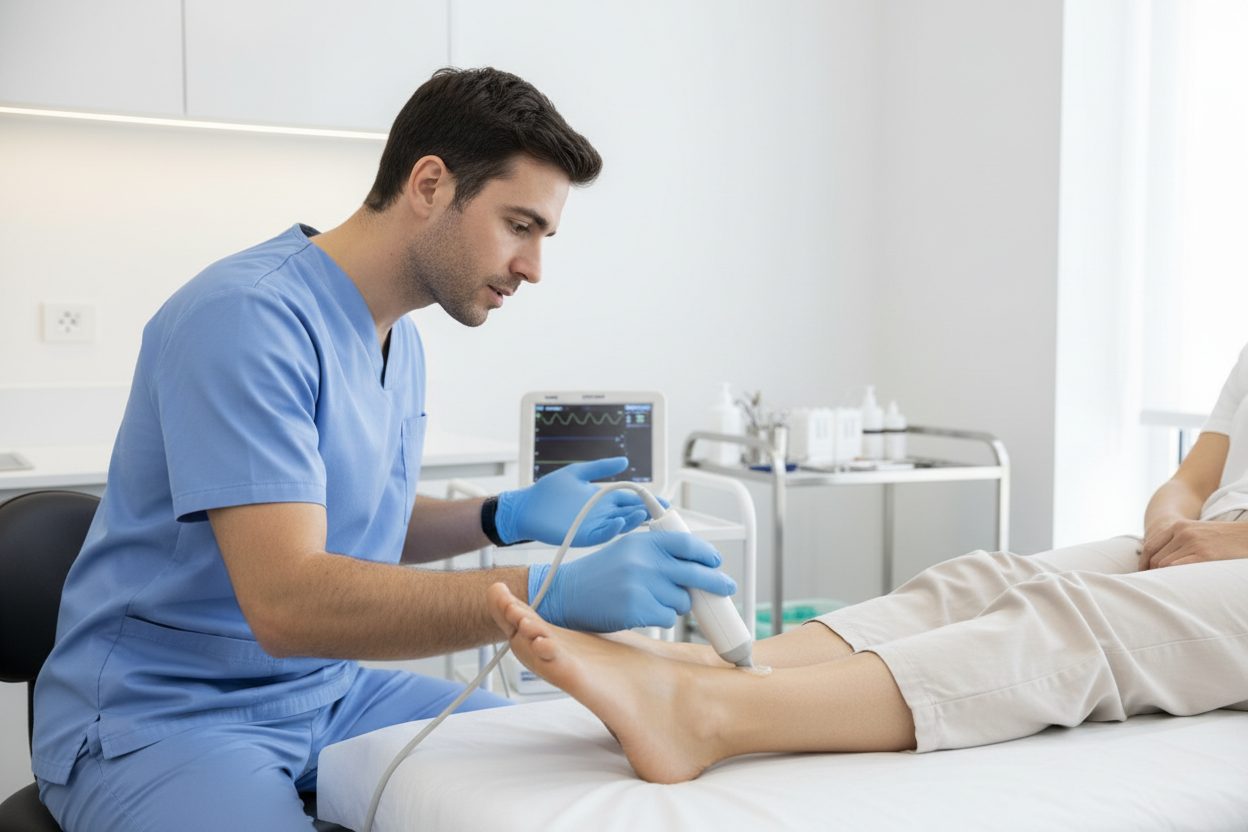
While compression therapy serves as the clinical cornerstone for managing chronic venous stasis ulcers, compression must never be applied blindly. Before prescribing therapeutic wraps or stockings, clinicians must measure the ankle-brachial index (ABI). This objective noninvasive test compares blood pressure in the ankles to the arms to assess arterial competence. If a patient receives high-pressure compression wraps while harboring undetected arterial blockage, the external pressure can completely cut off arterial supply, triggering rapid tissue necrosis and gangrene.
Arterial Leg Ulcers: When Restricted Flow Demands Protection
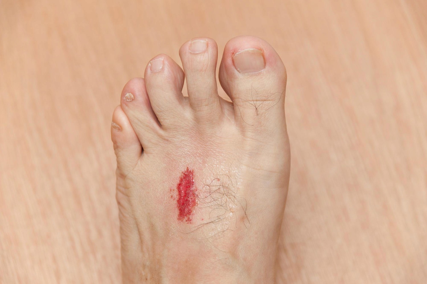
Arterial ulcers develop from the opposite circulatory defect: inadequate blood delivery to distal tissues. Caused by peripheral artery disease (PAD), atherosclerosis, or arterial vasospasm, these wounds emerge when narrowed vessels cannot supply enough oxygen and vital nutrients to meet metabolic demands. Because tissue cells are literally starving for oxygen, even trivial microtrauma—such as rubbing from a tight shoe—can trigger a persistent, deep wound.
Clinically, arterial ulcers present very differently from venous wounds. They typically appear on the outer ankle, top of the foot, or tips of the toes. They often feature distinct, "punched-out" circular margins, pale or gray wound bases, minimal drainage, and profound pain that tends to intensify when legs are elevated at night. The affected foot or leg may appear pale, shiny, hairless, and cool to the touch.
The management strategy for arterial ulcers diverges sharply from venous care. Compression is strictly contraindicated because external force further strangles compromised blood flow. Instead, the primary medical objective involves restoring vascular perfusion. Patients frequently require formal vascular evaluation for possible revascularization procedures, like angioplasty or surgical bypass, before the ulcer can safely begin healing. Until arterial blood supply improves, local wound management focuses on keeping the area dry, clean, and protected from bacterial colonization.
Diabetic and Neuropathic Ulcers: Relieving Unfelt Pressure
Diabetes creates a unique combination of risks for lower-extremity ulceration. Peripheral neuropathy blunts sensory awareness, meaning patients cannot feel friction, heat, cuts, or sustained pressure. Over time, motor neuropathy alters foot biomechanics, causing structural shifts that create focal high-pressure zones on the soles, heels, or toes. Autonomic neuropathy also dries out the skin, making it prone to cracking.
Evidence-based protocols from the IWGDF guidelines on diabetes-related foot disease emphasize that pressure relief through specialized offloading devices represents the foundational pillar of management. If mechanical shear and downward force continue striking the wound base, new cellular matrices are destroyed as fast as the body attempts to generate them. Offloading casts, specialized walking boots, and custom orthotics redistribute weight so vulnerable tissue can rest.
Managing diabetes-related ulcers also requires active control of blood glucose, aggressive screening for concurrent peripheral artery disease, and immediate treatment of localized infections. Without meticulous offloading and multidisciplinary attention, superficial skin breaks can rapidly extend deep into underlying tendon and bone structures.
Preparing the Wound Bed for Recovery
Regardless of whether an ulcer originates from venous congestion, arterial deficits, or neuropathy, chronic wounds frequently accumulate devitalized slough, hardened calluses, and bacterial biofilms. This nonviable material acts as a physical barrier that prevents healthy epithelial margins from migrating across the wound bed.
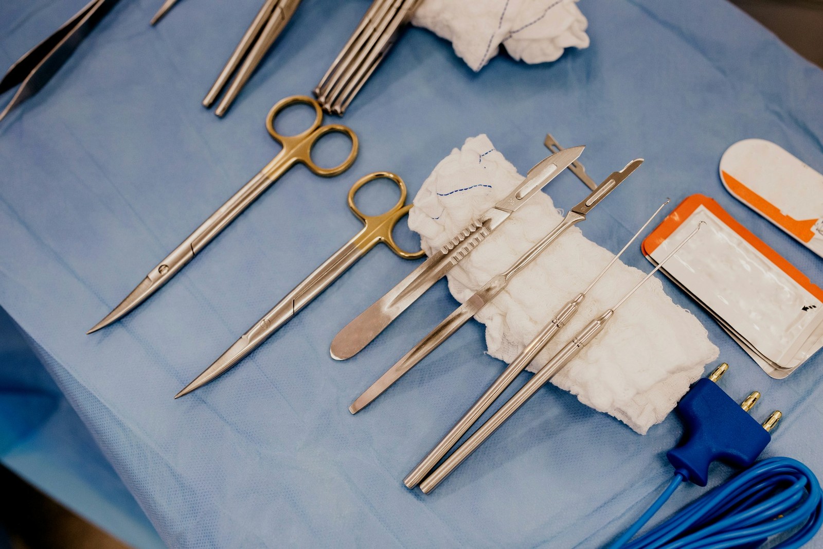
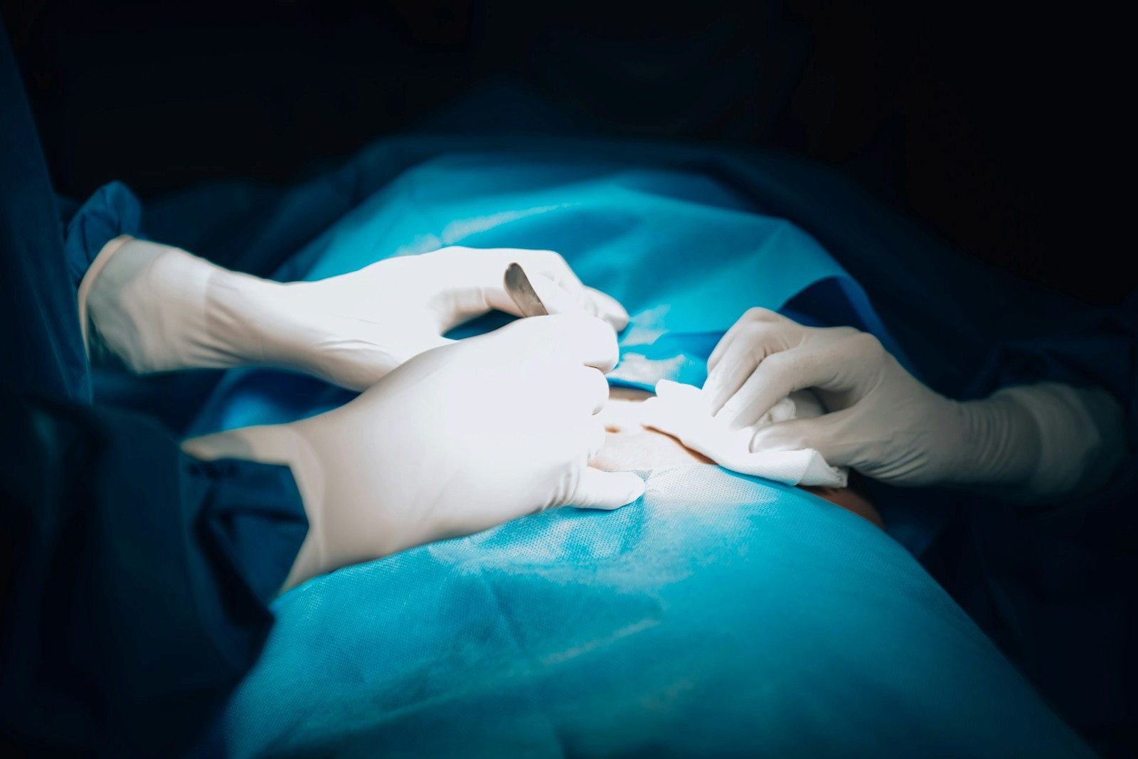
When clinically indicated, wound care clinicians perform targeted sharp debridement to carefully clear away nonviable tissue using sterile instruments. Removing devitalized matter stimulates biological signaling, transforming a stagnant ulcer into an active healing environment. Patients must never attempt self-debridement or cut calluses at home, as unsterile tools can introduce severe bacterial infections. In certain non-healing cases where severe tissue hypoxia resists standard interventions, providers may evaluate whether adjunctive hyperbaric oxygen therapy (HBOT) is appropriate. HBOT is not suitable for every wound and must be administered within a dedicated clinical hyperbaric chamber facility, rather than as a mobile or home-delivered therapy.
Warning Signs That Require Urgent Care
Lower-extremity ulcers can worsen rapidly if secondary infection develops. Contact your clinician or seek emergency care immediately if you notice spreading redness, warmth, red streaks moving up the calf, cloudy or foul drainage, fever, or sudden severe pain. Pale or blackened toes indicate critical vascular compromise requiring emergency medical intervention.
Comprehensive Clinical Support for Healing
Because accurate diagnosis governs every phase of care, patients should never manage non-healing ulcers alone. Anchor Wound Management provides individualized evaluations through clinic visits in Plano and Irving as well as mobile bedside care across Dallas–Fort Worth. If you need expert evaluation for a persistent wound, please reach out to schedule an evaluation with our team today.
Sources
- Robles-Tenorio A, Lev-Tov H, Ocampo-Candiani J. Venous Leg Ulcer (StatPearls). 2022.
- MedlinePlus Medical Encyclopedia. Venous insufficiency.
- International Working Group on the Diabetic Foot. IWGDF Guidelines on the prevention and management of diabetes-related foot disease (2023 update). 2023.