Conditions
Pressure Ulcer Treatment: How Care Changes With Wound Severity
Explore how pressure ulcer treatment changes across stages 1 to 4. Discover clinical dressing choices, repositioning tips, and when to seek urgent care.
Published · Anchor Wound Management

Overview
- Pressure injuries develop when sustained pressure and shear decrease blood flow over bony prominences, requiring different management strategies as tissue damage deepens.
- Early stages focus primarily on pressure redistribution, skin barrier protection, and routine position changes to prevent skin breakdown.
- Advanced stages involving full-thickness skin loss demand skilled clinical debridement, specialized moisture-balancing dressings, and targeted infection control.
- Accurate staging by a trained clinician ensures care plans adapt quickly to changing tissue characteristics, drainage levels, and nutritional needs.
When a loved one experiences skin breakdown from immobility, securing personalized pressure ulcer treatment is essential to protect healing tissue and prevent complications. These wounds, also called bedsores or pressure injuries, are not static. As damage extends from superficial skin into deeper fascia, clinical care shifts fundamentally. According to MedlinePlus guidance on pressure sores, these injuries occur when sustained pressure cuts off circulation to the skin, placing individuals confined to a bed or wheelchair at high risk. Understanding how wound characteristics evolve allows families and clinicians to implement targeted interventions. Comprehensive clinical management of pressure ulcers helps address both the surface wound and underlying forces causing cellular damage.
Mechanics of Pressure Injury Development
Pressure injuries arise when localized external forces compress vascular networks against internal bony structures. As detailed in a clinical review from StatPearls on pressure injury pathology, sustained pressure and shear compromise capillary perfusion, with the sacrum, ischial tuberosities, and greater trochanters accounting for approximately 70% of all reported injury locations. Collapsed capillaries deprive tissues of oxygen and micronutrients, causing metabolic waste accumulation, localized ischemia, and tissue necrosis.
Shear forces compound this injury when skeletal bone slides while overlying skin stays fixed against bed linens or chair cushions. This tension distorts microvessels, accelerating hypoxia. The National Pressure Injury Advisory Panel categorizes wounds into four numbered stages, unstageable wounds, and deep tissue injuries.
Adapting Pressure Ulcer Treatment Across Clinical Stages
Effective pressure ulcer treatment changes significantly as tissue loss deepens. Clinical goals must pivot from preventive skin preservation to bioburden control, dead tissue removal, and structural protection depending on anatomical involvement.
Stage 1: Intact Skin with Non-Blanchable Erythema
Stage 1 injuries present as localized areas of persistent redness that do not turn pale when pressed, known clinically as non-blanchable erythema. In darker skin tones, the area may present with persistent purple discoloration, warmth, coolness, or tissue firmness. The epidermal barrier remains intact without open skin breaks.
Treatment priorities center on immediate pressure relief and protecting the fragile epidermal layer. Caregivers must eliminate mechanical friction, implement strict turning schedules, and apply breathable barrier creams to defend against moisture. Massaging the reddened area is strictly contraindicated, as rubbing ischemic tissue causes secondary microvascular trauma. Protective transparent films or soft silicone dressings may also be used to reduce linen friction.
Stage 2: Partial-Thickness Loss of Skin
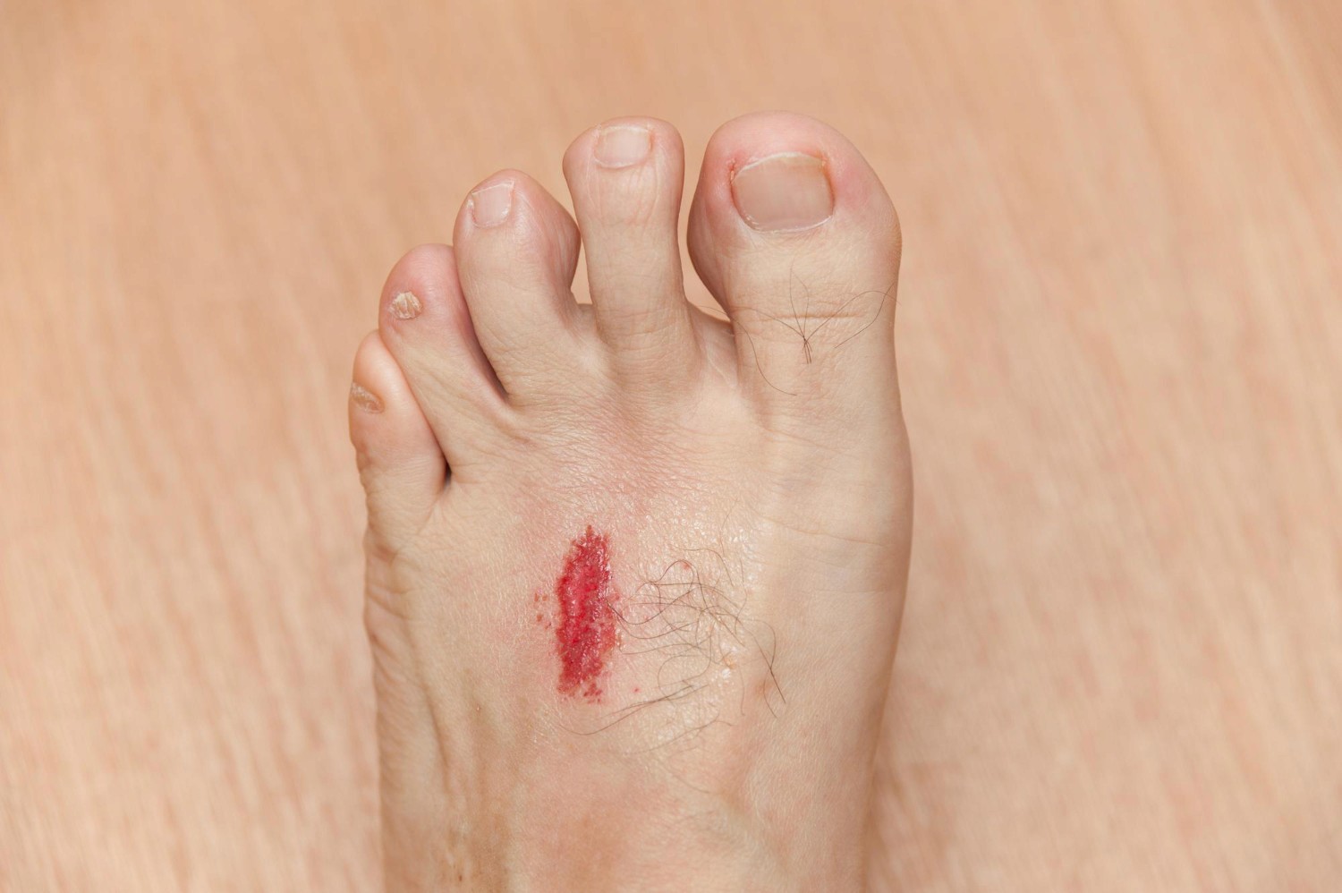
Stage 2 pressure injuries exhibit partial-thickness loss of the dermis. The wound presents clinically as a shallow, open ulcer with a viable, pink or red wound bed without slough or bruising. This stage also includes intact or ruptured serum-filled blisters caused by mechanical shear. Subcutaneous adipose tissue is not visible.
Care strategies focus on maintaining a balanced, moist healing environment while shielding exposed nerve endings. Cleansing is performed gently using sterile saline or non-cytotoxic cleansers. Clinicians commonly prescribe hydrogels, hydrocolloids, or thin silicone foam pads to absorb light exudate while preventing dressing adherence. Gentle handling during dressing changes is critical to minimize trauma.
Stage 3: Full-Thickness Skin Loss
Stage 3 represents full-thickness tissue destruction where subcutaneous fat becomes visible within the open ulcer cavity. Deeper structures like bone, tendon, and muscle remain safely covered. Slough or eschar may be present, and undermining or tunneling beneath intact skin margins often begins to develop.
Care priorities shift toward managing wound drainage, addressing dead tissue, and eliminating dead space. Clinicians evaluate whether sharp, enzymatic, or autolytic debridement is appropriate to clear necrotic material harboring bacterial biofilms. Alginate or hydrofiber dressings are packed lightly into wound cavities to absorb drainage, while secondary border dressings maintain protection. Systemic nutritional support becomes vital, as tissue repair requires elevated protein intake.
Stage 4: Full-Thickness Skin and Tissue Loss
Stage 4 injuries involve catastrophic tissue loss extending past subcutaneous layers into palpable or directly visible fascia, muscle, tendon, ligament, cartilage, or bone. Slough, black necrotic eschar, and extensive undermining or sinus tracts are frequently observed. Because bone is exposed or directly palpable, the risk of osteomyelitis and systemic infection escalates sharply.
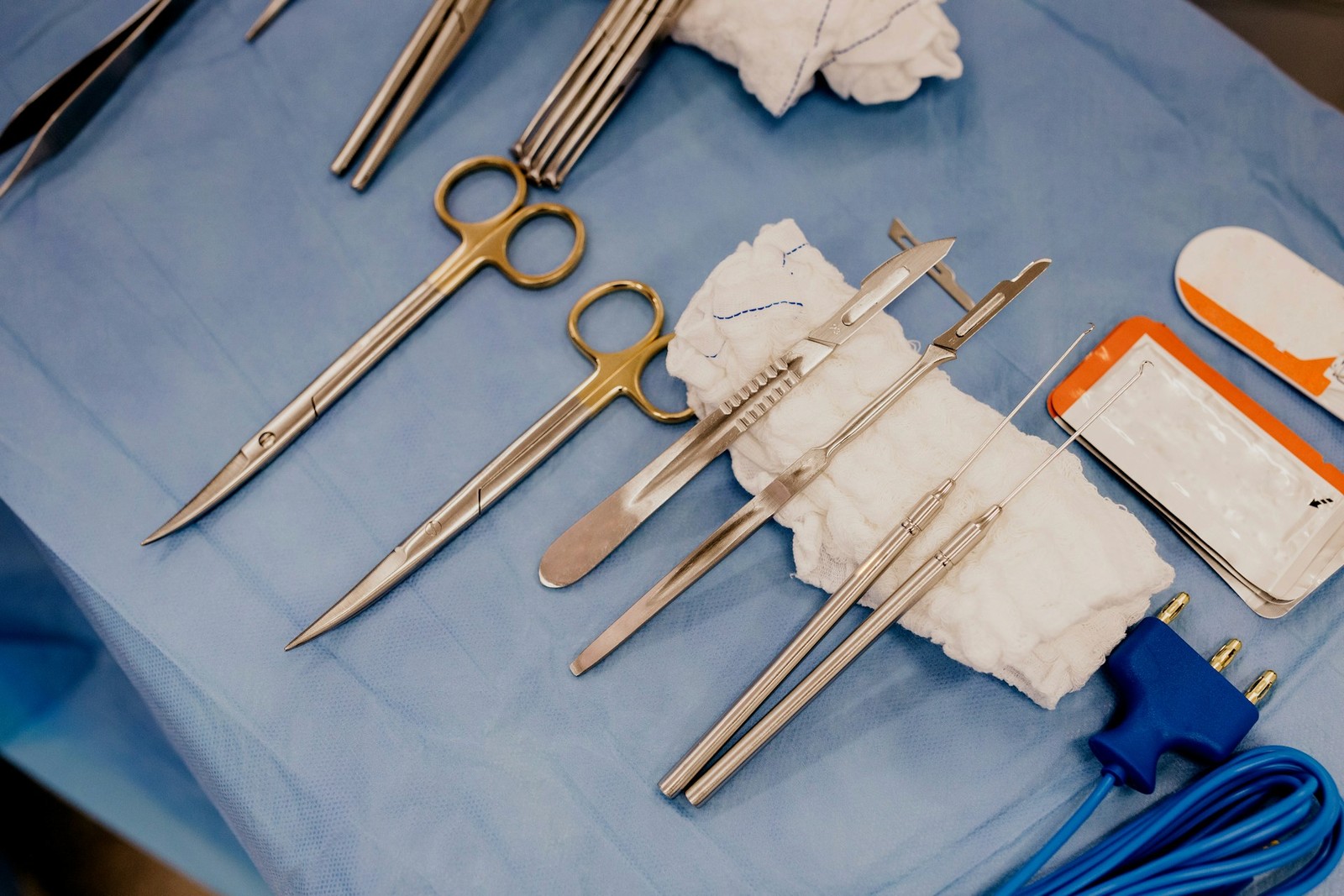
Management demands aggressive medical care. Specialized surgical or bedside debridement is necessary to excise extensive nonviable tissue. Antimicrobial dressings, such as silver-impregnated ropes or cadexomer iodine, help suppress bacterial burdens. Deep cavities require loose packing to prevent premature surface closure over unresolved infections. Advanced therapies, such as negative pressure wound therapy, may be utilized under medical supervision to draw wound edges together.
Deep Tissue Pressure Injuries and Unstageable Wounds
Two complex classifications require special caution during evaluation: deep tissue pressure injuries and unstageable wounds. A deep tissue injury presents as intact or non-intact skin exhibiting persistent, localized deep red, maroon, or purple discoloration, or a blood blister resulting from intense shear at the bone-muscle interface. These areas can deteriorate rapidly into full-thickness ulcers despite appearing stable initially.
Unstageable wounds occur when the true depth of tissue destruction is completely obscured by slough or thick eschar. Until nonviable tissue is debrided to expose the base, the wound cannot be staged. However, stable, dry, intact eschar on an ischemic heel without erythema is generally left intact as a biological cover unless infection develops.
Fundamental Support Across Every Healing Phase
Regardless of staging, successful healing depends heavily on systemic and environmental factors. Repositioning individuals every two hours in bed, and every fifteen minutes in wheelchairs, reduces capillary compression. Specialized support surfaces—such as alternating-pressure mattresses or foam cushions—disperse body weight. Following structured home wound care safety guidelines ensures that family members maintain clean technique and reduce friction during transfers.
Adequate hydration and clinical nutrition provide essential building blocks for tissue regeneration. Dietitians often recommend targeted protein supplementation, vitamin C, and zinc to support collagen synthesis. Furthermore, maintaining meticulous skin hygiene by keeping periwound skin dry and free from maceration prevents secondary breakdown.
Warning Signs Requiring Urgent Medical Evaluation
Pressure injuries can deteriorate swiftly if bacterial colonization transforms into an invasive infection. Patients and caregivers should monitor wounds daily and seek immediate clinical attention if any of the following warning signs arise:
- Spreading redness, warmth, or swelling expanding outward from the wound perimeter.
- A sudden increase in drainage, cloudy discharge, or foul odor emerging after gentle cleansing.
- Systemic infection signs, including chills, confusion, or a fever exceeding 100.4 degrees Fahrenheit.
- New or escalating localized pain that persists despite scheduled comfort measures.
- The appearance of blackened tissue, rapid wound cavity enlargement, or visible bone.
Partnering with Specialized Wound Care Clinicians
Navigating the complexities of pressure injuries requires ongoing assessment from experienced clinicians. At Anchor Wound Management, our team provides clinical visits and convenient mobile bedside wound care across the Dallas–Fort Worth region, bringing expert staging, customized dressing regimens, and advanced debridement directly to patients. With offices in Plano and Irving, our clinicians work closely with families to coordinate healing. If you or a family member faces skin breakdown, do not wait for complications to escalate. Please contact Anchor Wound Management to schedule an evaluation and recovery plan.
Sources
- Zaidi SRH, El Khatib S, Sharma S. Pressure Injury (StatPearls). 2026.
- MedlinePlus, U.S. National Library of Medicine. Pressure Sores.